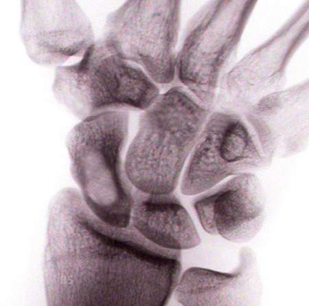
Image: Enchondroma in the scaphoid bone (scaphoid cyst), bright spot in the large scaphoid carpal bone. This localization on the wrist is less common.
Enchondroma is a benign tumor of the bone and consists of cartilage. In principle, it can occur on any bone. The exact cause of its occurrence is unclear. This cystic bone tumor can occur individually or in large numbers. The latter variant is referred to as enchondromatosis.
The enchondroma is clearly visible as a dark spot on the X-ray, possibly with calcifications, so that no further diagnosis is usually necessary.
What symptoms are typical of enchondroma?
Enchondromas rarely cause direct symptoms. It is therefore often detected as an incidental finding on an X-ray. However, a larger enchondroma can become visible through swelling. Furthermore, the outer layer of bone, the cortical bone, can be so thinned that it can break or fracture completely in the case of minor injuries (so-called pathological fracture). In this way, the enchondroma makes itself felt through pain, as in a bone fracture. An enchondroma should also be considered, for example, in cases of unclear wrist pain. In the case of an uncomplicated fracture, it is advisable to remove the enchondroma only after the fracture has healed. However, if the fracture requires surgery, this is combined with the removal of the enchondroma.
Facts and statistics
The following facts are known about this benign bone tumor:
- It is the most common tumor on the skeleton of the hand.
- The enchondroma can degenerate. However, this is extremely rare in the hand.
- 90% of all bone tumors in the hand are enchondromas.
- In 35% of cases, the tumor develops on the hand.
- People between the ages of 10 and 40 are usually affected.
- Enchondroma is less common on the carpal bones than on the long bones of the hand.
What should you know about enchondroma surgery?
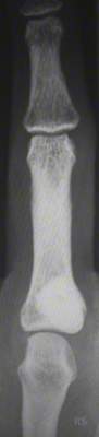
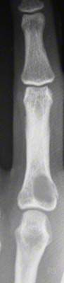
Image: Left: Enchondroma at the base of the proximal phalanx with cortical thinning; right: filling of the defect with hydroxyapatite ceramic.
When should surgery be performed?
- A small enchondroma that does not cause any symptoms can be observed (X-ray examinations).
- If you are not sure whether it is an enchondroma, further diagnostic measures such as CT or MRI can be helpful.
- Large enchondromas are also operated on without symptoms to prevent a hernia.
- A small enchondroma should also be operated on if symptoms occur.
The operation
Enchondroma surgery consists of complete curettage of the tumor. The approach can be on the extensor or lateral side of the finger. The bone is exposed and opened – e.g. by milling or chiseling – while protecting the important structures. The glassy-cartilaginous enchondroma is then removed with a curette. This must be done completely. If remnants remain, recurrence is inevitable.
The resulting bone cavity is then filled. Depending on the size, this can be done with cancellous bone (bone marrow) from the pelvic bone or from the radius (forearm bone). Some hand surgeons also use chemically processed bone material from the cadaver (allograft)6. There are also bone substitutes made of hydroxyapatite that can be used. The latter saves a second wound and the sometimes not insignificant pain after removing bone from the pelvis. However, it is not absolutely necessary to fill the cavity if the bone wall is stable.5 The cavity will be refilled by the body over time.
The following should be noted after enchondroma surgery.
The follow-up treatment depends on the extent of the findings and the associated operation. Therefore, immobilization can range from a few days to 4-6 weeks for fractures. The movement exercises and the start of physiotherapy also vary depending on the operation.
Even after healing, further X-ray examinations should be carried out, as the enchondroma can recur in approx. 4.5% of cases.7
Here are some pictures taken during the operation with the image converter:
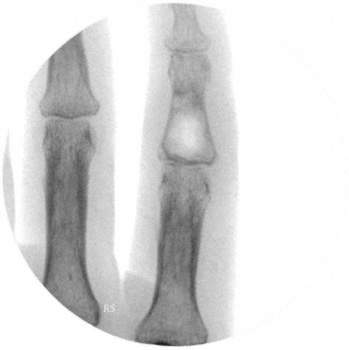
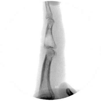
- 1.2 Enchondroma of the proximal phalanx of the index finger (X-ray in 2 planes). The cortical bone (stable outer bone wall) is visibly thinned.
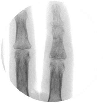
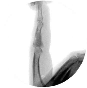
- 3.4 Defect filled with cancellous bone from the iliac crest