The metacarpal bones, or MHK for short, are called ossa metacarpalia.
All metacarpal bones belong to the small tubular bones. They can be divided into a shaft, neck = collum (between the head and shaft), a spherical head (caput) and a base. The metacarpal base is connected to the carpal bones via articular surfaces (carpometacarpal joints). The head is connected to the metacarpophalangeal joints (metacarpophalangeal joints) and the metacarpophalangeal joints (metacarpophalangeal joints) and the metacarpal bones are easily palpable on the extensor side of the metacarpus from the base to the head.
First metacarpal bone – MHK I
The first metacarpal bone is the strongest, but the shortest of all. At the base, the articular surface is saddle-shaped and forms the thumb-saddle joint with the large polygonal bone = os trapezium.
Osteoarthritis in this joint is known as thumb saddle joint osteoarthritis = rhizarthrosis. The disease leads to a narrowing of the joint space and even destruction and stiffening of the joint.
2nd-5th metacarpal bone
The following images show the metacarpal bones of the left hand in the order MHK II, III, IV and V.
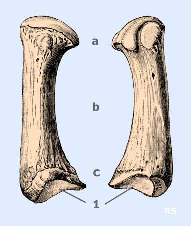
MHK II:
The Os metacarpale II is the longest of the 5 bones.
The base of the 1st metacarpal is the insertion of the tendon of the longextensor carpi radialis longus (wrist extensormuscle)
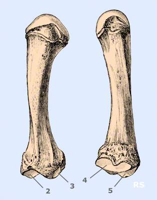
- 2, articular surface of the small polygonal bone (Os trapezoideum)
- 3, articular surface of the large polygonal bone (Os trapezium)
- 4, articular surface to the 3rd metacarpal bone
- 5, = 2
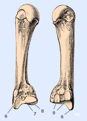
MHK III:
- 6, styloid process for the insertion of the tendon of the small wrist extensor (extensor carpi radialis brevis muscle)
- 7, articular surface to the 2nd metacarpal bone
- 8, articular surface to the capitate bone (Os capitatum)
- 9, articular surface to MHK IV
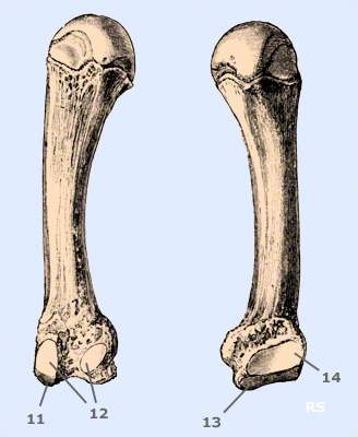
MHK IV:
- 11, articular surface to the capitate bone (Os capitatum)
- 12, articular surface to the 3rd metacarpal bone
- 13, articular surface to the hook bone (Os hamatum)
- 14, articular surface to MHK V
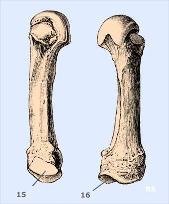
Os metacarpale V:
- 15, articular surface to the 4th metacarpal bone
- 16, articular surface to the hook bone (Os hamatum)
In many cases, an intramedullary K-wire osteosynthesis can be performed for displaced fractures of the MHK V neck (MHK V collum fracture). This minimally invasive operation only requires a small incision in the area of the base. The bone is drilled out, 2 wires are placed in the medullary cavity and advanced into the head. These are stretched against each other and enable good stabilization. This technique can also be used for collum fractures of the 4th metacarpal.