Basal cell carcinoma is also known as basal cell carcinoma and basal cell epithelioma. As the name suggests, it develops from the basal cell layer in the skin. It can be divided into further subtypes by the pathologist:
- Trunk skin basal cell carcinoma (superficial basal cell carcinoma) usually grows superficially.
- Pigmented basal cell carcinoma: It is visually distinguished from the others by its brown color.
- Nodular (nodular) basal cell carcinoma
- sclerodermiform basal cell carcinoma
- Other and mixed forms
Basal cell carcinoma is also often referred to as white or light skin cancer – which can be rather misleading.
It grows destructively but does not usually spread (metastasize). If left untreated, it destroys important structures, particularly in the head and neck area, and can therefore lead to death. If superficial structures are destroyed, it is called a rodenous ulcer, if deep structures are affected, it is called a terebranous ulcer.
How does basal cell carcinoma develop?
Basal cell carcinoma mainly develops in connection with increased UV radiation from the sun or solarium. It particularly affects the light Nordic skin type, which is characterized by: pale skin, blue eyes, blonde or red hair. Such people primarily turn red instead of brown when exposed to sunlight.
However, genetic factors also play an important role. Basal cell carcinoma tends to run in families.
Arsenic and treatment with drugs that weaken the immune system (immunosuppressants) can also promote light skin cancer.
Statistics
- White skin cancer occurs in 80% of cases in the head/neck area.
- Spreading (metastasizing) is extremely rare and has only been observed in isolated cases.
- In Australia, basal cell carcinoma is the most common form of cancer.
- In Germany, around 100 out of every 100,000 people fall ill every year.
- The average age of those affected is 60, although light skin cancer can also occur in much younger people.
How do I recognize a basal cell carcinoma?
White skin cancer can look very different depending on the subtype and stage. Some forms (trunk skin basal cell carcinoma) are characterized only by redness that does not go away. Sclerodermiform basal cell carcinoma is often characterized by redness with scaling and can be mistaken for eczema.
A typical feature is often the so-called pearl cord border that forms the edge. Overall, the basal cell carcinoma often appears as a crater-like skin change that oozes centrally.
The areas of the body that are particularly exposed to the sun are typical for the localization: Head, neck, shoulders, lower legs.
The advanced stage is characterized by defects and destruction. Sometimes the nose, eyes, midface and other areas have to be removed as part of the operation.
The diagnosis is made by the plastic surgeon based on the appearance.
To get a small impression of the many appearances of this type of skin cancer, click on basal cell carcinoma pictures.
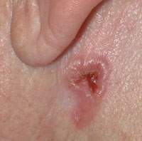
Picture: Typical basal cell carcinoma at the angle of the jaw with pearl cord, crater formation and encrustation.
Should basal cell carcinoma removal be performed surgically?
The standard treatment for basal cell carcinoma is removal by surgery at the Yuveo Clinic Düsseldorf and assessment under the microscope by the cooperating pathologist.2
In addition to surgery, however, there are a number of other treatment options:
- Radiotherapy: This is suitable for inoperable basal cell carcinoma or inoperable patients.
- Curettage: Can be successful with superficial skin cancer of this form. However, it is not possible for the pathologist to check the edge of the incision.
- Cryotherapy: The basal cell carcinoma is effectively killed with cold. The procedure is suitable for small, superficial basal cell carcinoma and older patients with a high risk of surgery.
- Laser therapy
- Photodynamic therapy: In this method, which can be used for superficial basal cell carcinoma, the basal cell carcinoma cells are first made light-sensitive with a special cream (active ingredient: delta-aminolevulinic acid) and then destroyed with so-called Wood light.
- Treatment with the drugs Imiquimod and 5-Fluorouracil only for superficial basal cell carcinoma.
What all these procedures have in common is the fact and the disadvantage that no microscopic examination can be carried out. It is therefore not possible to know whether the cancer has been destroyed down to the healthy tissue. Furthermore, after surface therapy (laser, curettage = scraping, ointments), problem areas can develop that continue to grow in depth.
How is basal cell carcinoma surgery performed?
The aim of the operation is to completely remove the tumor. If this is confirmed by the pathological examination, the patient is considered cured.
Surgery at our clinic in Düsseldorf can often be performed under local anesthesia and on an outpatient basis as long as no deep layers are affected and the size does not exceed a certain level.
The surgical principle consists of basal cell carcinoma removal with a safety margin and subsequent microscopic examination. The safety margin should also be 3-5 mm for smaller basal cell carcinomas – and even more for larger ones.
The removed basal cell carcinoma is marked with a thread to provide orientation. If the pathologist still sees extensions in certain places or a safety margin that is too small, he can inform the plastic surgeon.
One- or two-stage procedure?
In principle, both procedures can be chosen. However, in the case of a large basal cell carcinoma, unfavorable localization or uncertainty when assessing the margins, the defect should not be closed until the pathologist can confirm the removal with a sufficient safety margin.
This means that the resulting defect is first sterilely dressed or covered with replacement skin until the wound is finally closed.
In the single-stage procedure, the defect is closed immediately. This is an adequate method for small and medium-sized stages, if the closure is not surgically complex.
Defect cover
Cutting out the basal cell carcinoma is not the art, but the subsequent defect coverage. Plastic surgeons and oral and maxillofacial surgeons have different procedures at their disposal.
In the simplest case, the defect is covered by moving the skin over the defect after detaching the skin from the underlying tissue.
The most important form of defect coverage is the so-called sliding and pivoting flap. For slightly larger defects, a skin flap is moved into the defect using various incision patterns. (See also V-Y-plasty)
The option of skin grafting should also not be forgotten for skin cancer surgery. Defect coverage is achieved by inserting a skin graft from another area with similar quality and excess skin into the defect. The graft must now heal here.
In the case of very large defects, the range extends from large swing flaps to so-called free flaps. Often the nose or eye socket must also be reconstructed or favorable conditions created that provide good conditions for the epithesis. An epithesis is, for example, an artificial nose or an artificial ear that can be attached.
What are the recommendations for post-operative care?
No matter how the treatment was carried out: Follow-up care in the form of annual check-ups for at least 3 years is recommended by our team in Düsseldorf. We also encourage our patients to perform self-examinations.
What precautions should you take?
Basal cell carcinoma prevention is based on 3 pillars:
- Skin examination: independently or by the partner
- Skin cancer screening: preventive examinations at the dermatologist
- Sun protection: avoid direct sunlight, use high sun protection factor sun creams, seek shade, wear a hat and clothing
Pubmed:
- 1, Guidelines for basal cell carcinoma management:
Guidelines for the management of basal cell carcinoma – Telfer NR, Colver GB, Morton CA; British Association of Dermatologists. – Br J Dermatol. 2008 Jul;159(1):35-48. review . - 2, Interventions in basal cell carcinoma of the skin:
Interventions for basal cell carcinoma of the skin. – Bath-Hextall FJ, Perkins W, Bong J, Williams HC. – Cochrane Database Syst Rev. 2007 Jan 24;(1):CD003412. Review . - 3, Management of basal cell carcinoma in adults Clinical practice guidelines. – Dandurand M, Petit T, Martel P, Guillot B; ANAES. – Eur J Dermatol. 2006 Jul-Aug;16(4):394-401 .
Other:
- 4, AWMF Guideline – Basal cell carcinoma – Register No. 032/021 – Development stage 1 – Status 09/2007 – next update planned 4/2010