On this page you have the opportunity to see the different ‘basal cell carcinoma faces’ so that you can better assess and recognize skin changes in yourself and in acquaintances, relatives or friends.
However, this small collection of pictures is no substitute for a medical degree. You should consult a (skin) doctor for any unusual skin changes!
For more information on the disease, symptoms, treatment and surgery, click on: Basal cell carcinoma or white skin cancer.
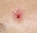
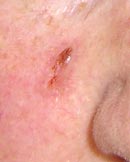
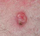
We start with the classic basal cell carcinoma. In the center you can see an ulceration (= open area, ulcer) with a bloody crust and the typical border, also known as the pearl cord border.
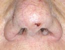
This is a large nodular basal cell carcinoma with a blood crust on the lateral nasal roof.
The small blood vessel dilatations on the nodular change in this image are also typical of diene cancer.
Small vascular dilatations and a crust are also visible here. On closer inspection, it is clear that the crust only makes up a quarter of the skin cancer. The basal cell carcinoma extends down to the middle of the upper lip.
The light-colored skin cancer on the tip of the nose is in an unfavorable position for wound closure. However, a skin graft can be used here. Even in this picture, the crust only makes up part of the pathological change.
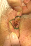
This skin cancer is also a basal cell carcinoma. The marking shows a safety distance of 4 mm as seen with magnifying glasses.
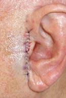
Right: After removal of the basal cell carcinoma, the defect was closed by detaching the skin (= stretch plasty) and moving it in a horizontal direction.
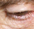
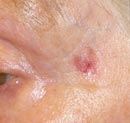
This case is somewhat more ‘insidious’ than those described above. It is simply a red spot about 3 mm wide with a discreet indentation. You can see it in the center at the lower edge of the eyelid.
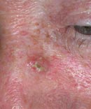
Another basal cell carcinoma on the lateral bridge of the nose. The patient initially noticed scaling and redness here, as with eczema. However, the change did not disappear and slowly became larger.
This is a similar-looking white skin cancer to the one described above, but much larger.
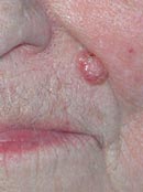
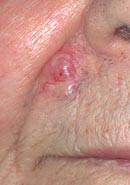
A nodular basal cell carcinoma in the nasolabial fold. With this size and localization, the basal cell carcinoma can be easily removed. The scar is then barely visible in the nasolabial fold.
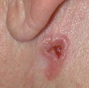
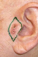
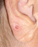
Here you can see a basal cell carcinoma that has already affected 1/3 of the auricle.
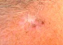
This skin cancer appears quite small. On closer inspection, however, a slightly lighter border around the encrustation is noticeable. This brightening is also part of the cancer.
The patient complained of an eczema-like change that repeatedly flaked. After scratching, bleeding and crusting occurred. This was also a basal cell carcinoma.
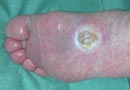
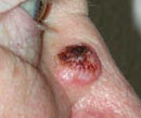
Many years ago, an elderly patient had a basal cell carcinoma (very rare on the sole of the foot) irradiated. The radiation damage has now led to the death of the surrounding tissue down to the flexor tendons of the foot.
This relatively poorly identifiable structure in the middle of the picture is also a white skin cancer. It is almost as large as the nostril visible in the picture.
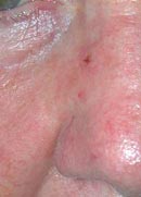
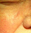
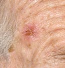
This basal cell carcinoma on the temple is only noticeable due to an irregularly reddened area.
This is a relapse. The white area is the scar left after the operation about 3 years ago. The slight redness on the lower and right edges corresponds to the basal cell carcinoma that has grown again.
Another basal cell carcinoma with crusting and rim on the cheek.
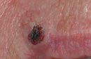
Is this a basal cell carcinoma?
Yes, it can also look like this. It is a rare pigmented basal cell carcinoma, which can easily be mistaken for a mole. For this reason, it is questionable to call it white skin cancer.
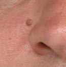
Nodular basal cell carcinoma with a small crust.
This finding looks like an almost healed skin abrasion. In fact, however, this is also a “light” skin cancer.